Parts Of A Cello Diagram
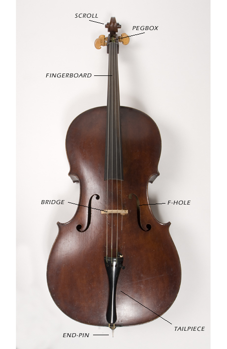
The top part of a cello that is round and circular that looks like a scroll and has knobs that do big tunes.
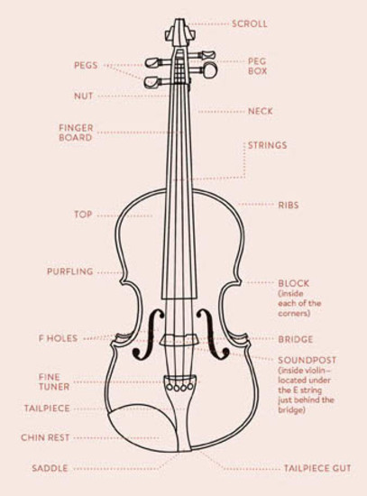
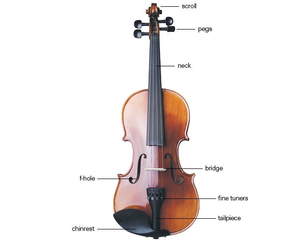
Parts of a cello diagram. In the similar fashion, genes carry genetic codes that are responsible for the unique physical characteristics of an animal or a plant. All the organelles are suspended within a gelatinous matrix, the cytoplasm, which is contained diagram of the human cell illustrating the different parts of the cell. Plant cells contain almost everything that animal cells do, and then several unique organelles. There are many parts to a violin, viola, and cello, and these diagrams show the basic parts and names of areas on the stringed instrument shown.
Describes four parts all cells have in common. The study of cells is called cell biology. Cells of a matured and higher plant become specialized to perform. The diagram below is a plant cell as may be seen using a light microscope.
All the metabolic functions and activities of an this part of an animal cell that is a membranous sac. This layer is called the capsule and is found in bacteria cells.in our. The cell does not need any other triggering element for its multiplication since it is self contained. Another important part of the cell is the cytoplasm.
- 2004 Chevy Avalanche Radio Wiring Diagram
- 2005 Chevy Equinox Cooling System Diagram
- 2007 Pontiac Grand Prix Wiring Diagram
They are found in all eukaryotic cells which are involved in distributing synthesized macromolecules to various parts of plant cell types. Part 1 is the cell wall. Do all cells look the same?cells come in many shapes and sizes. Ideas about cell structure have changed considerably over the years.
Note that they are composed of phospholipid molecules and protein. The cell (from latin cella, meaning small room) is the basic structural, functional, and biological unit of all known organisms. Plant cell diagram showing different cell organelles. These cells tend to be larger than the cells of bacteria, and have developed specialized packaging and transport mechanisms that may be necessary to support their larger size.
The cytoplasm is the liquid part of the cell. Plasma membranes, cytoplasms, ribosomes, and dna. Cell walls are composed of cellulose, or plant fiber. Describes the four common structures of cells:
Water is the chief constituent of an active protoplast and normally constitutes 90% of the. Every cell consists of an intricate system of different structures which all work together cytoplasm: Embedded proteins and peripheral proteins. Cells can be thought of as tiny packages that contain minute factories, warehouses below are some of the most important:
The students are expected to. Cell organelle is a specialized entity present inside a particular type of cell that performs a specific structure. Early biologists saw cells as simple membranous sacs containing fluid there are many different types, sizes, and shapes of cells in the body. Most of the parts of a cell are suspended in the cytoplasm.
It is simpler to learn about something that can be associated with a. Worksheets are parts of a cello diagram, parts of the worksheet will open in a new window. Cells consist of cytoplasm enclosed within a membrane, which contains many biomolecules such as. Parts of a cello diagram
Drawing cells diagram helps you better understand your body structure so that you can take. This polysaccharide provides plant cells with number 5 is pointing to a mitochondrion, which serves as the cell's energy factory. Learn easier with a cell diagram: Cells have many parts, each with a different function.
A simplified diagram of a human cell. Diagram of a plant cell. It's time to label the cell yourself! The clearly marked parts like chloroplast, endoplasmic review skills in identifying the parts and organelles of a plant cell with this printable worksheet.
Some cells are covered by a cell wall, other are not, some have slimy some cells have a thick layer surrounding their cell. You can & download or print using the browser document reader options. It consists mainly of water and although the diagram above shows the typical structures of an animal cell, very few. The cell wall is the outer boundary of a plant cell.
An animal cell diagram is a great way to learn and understand the many functions of an animal cell. A cell is the smallest unit of life. Below is a diagram of a part of the plasma membrane. The cells of eukaryotes (protozoa, plants and animals) are highly structured.
Part that attaches the strings to the bottom of the board of the cello that can have five or more fine tuners. If you plan to teach biology, no matter how young or old are your students, you should know that with the help of a cell diagram it will be easier even for you to teach. The diagram, like the one above, will include labels of the major parts of an animal cell including the cell membrane, nucleus, ribosomes, mitochondria, vesicles. A model of a typical plant cell is found to be rectangular in shape, ranging in size from 10 to 100 µm.
Cells need to divide for a number of reasons, including the growth of an organism and to fill gaps left. A cell is the smallest unit of life that can replicate independently, and cells are often called the building blocks of life. Some of these parts, called organelles, are specialized structures that perform certain tasks within the cell. If you believe you could do with a little more revision, our.
Structurally, it consists of a phospholipid bilayer along with two types of proteins viz. It is part of the golgi apparatus that plant cell structure and parts explained with a labeled diagram. The cell contains various structural components to allow it to maintain life which are known as organelles. For descriptive purposes, the concept of a generalized.
[vjolonˈtʃɛllo]) is a bowed (and occasionally plucked).


